Animal Housing Platform

SPF Animal Facility

SPF Animal Facility

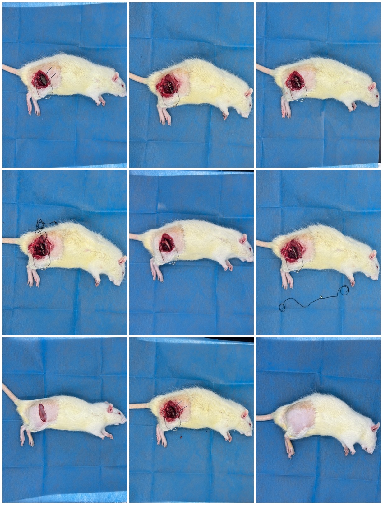
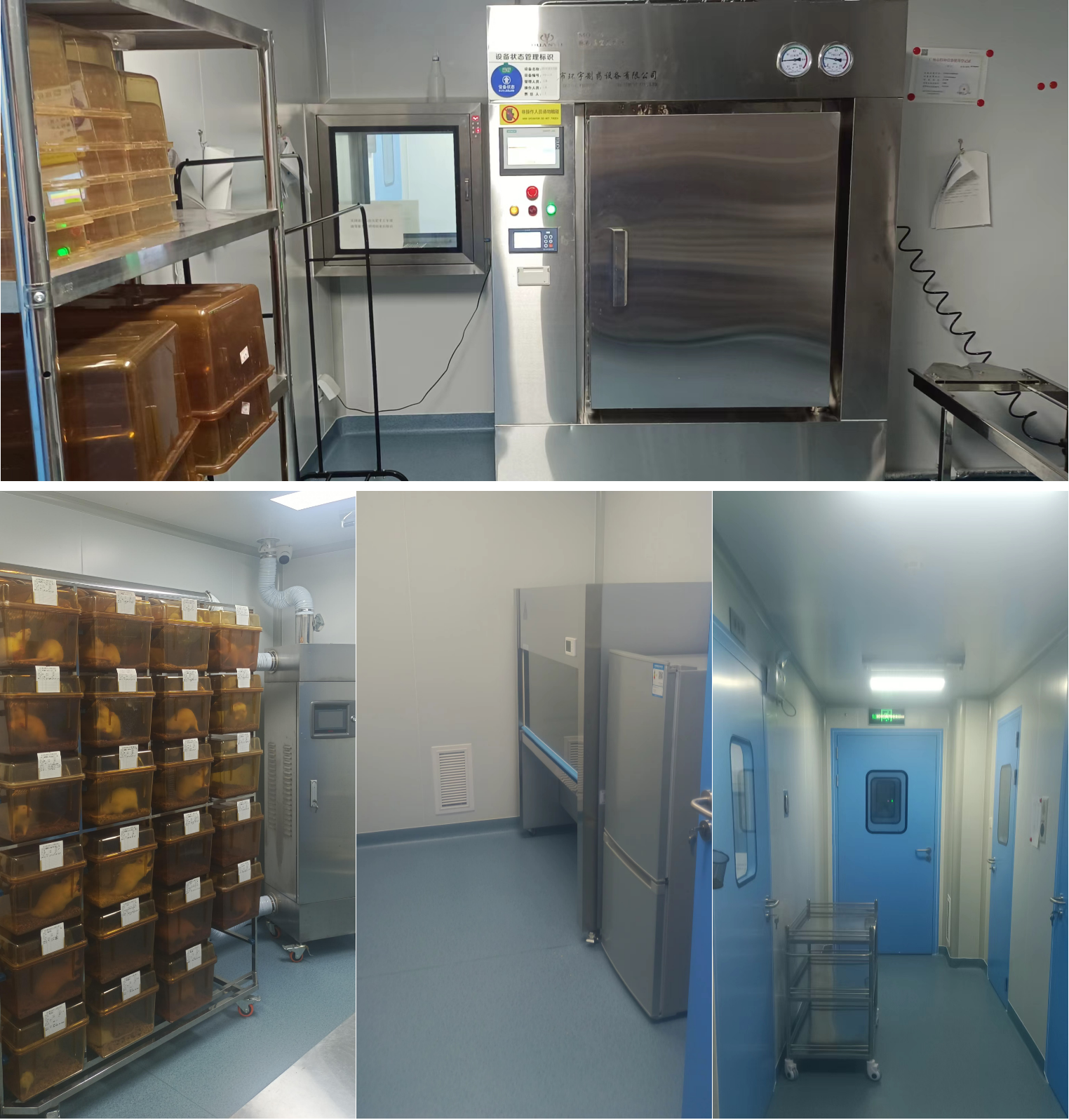
Located in Panyu Shilou Innovation Technology Park, equipped with 500+ mouse IVC cages and 150 rat IVC cages. The barrier system includes independent animal laboratories for surgical operations and behavioral testing.

Standardized feeding procedures and environmental monitoring ensure animal health and data stability.

Neurological Models

CSR Model

CSR Cervical Radiculopathy Model

Key Points

By exposing the C7-T1 lamina and inserting a polyamide compression line through the spinal canal at the C5-T1 nerve roots, sustained compression is achieved to replicate cervical radiculopathy.

Assessment & Duration

Gait scoring, gait pattern analysis, MRI positioning, and HE staining. Duration: 7-14 days.

MCAO Model

MCAO Middle Cerebral Artery Occlusion Model

Key Points

Exposing the common, external, and internal carotid arteries, inserting a filament through the internal carotid to temporarily occlude the middle cerebral artery, simulating ischemic injury.

Assessment & Duration

TTC staining, Longa scoring for infarct size and neurological deficit assessment. Duration: ~3 days.

Musculoskeletal Models

KOA Model

KOA Knee Osteoarthritis Model

Key Points

Intra-articular injection of type II collagenase solution to induce cartilage degeneration and inflammation, forming knee osteoarthritis-like pathology.

Assessment & Duration

MicroCT, HE and Safranin O/Fast Green staining for structural and pathological assessment. Duration: 35-42 days.

ACLT Model

ACLT Anterior Cruciate Ligament Transection Model

Key Points

Using inhalation anesthesia, medial knee approach to expose and transect the ACL, confirmed by Lachman test, establishing secondary osteoarthritis model.

Assessment & Duration

MicroCT, HE and Safranin O/Fast Green staining for bone and cartilage damage assessment. Duration: 28-56 days.

Bone Defect Model

Rat Femoral Bone Defect Model

Key Points

After anesthesia, blunt dissection between the vastus lateralis and biceps femoris exposes the femur. After fixation with a stainless steel plate, a 4mm bone segment is resected using an osteotomy guide and wire saw. PMMA bone cement is placed in the defect area.

Assessment & Duration

MicroCT 3D reconstruction, HE staining, Masson staining, and biomechanical testing. Duration: typically 4-12 weeks depending on observation timepoints.